SSM: A New Model for ob/ob Mouse Fat Study
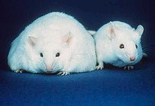
Ob/ob Mouse
|
||
Why need SSM?
|
||
What's SSMSSM=Segmental Shape Model SSM is a novel curve deforming model with the capacity of partially control the shape similarities. It provide a powerful tool for medical segmentation and other applications. Here we applied it in the fat study for ob/ob mouse.
|
||
Method for ob/ob mouse fat analysis
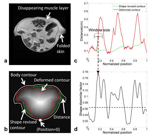
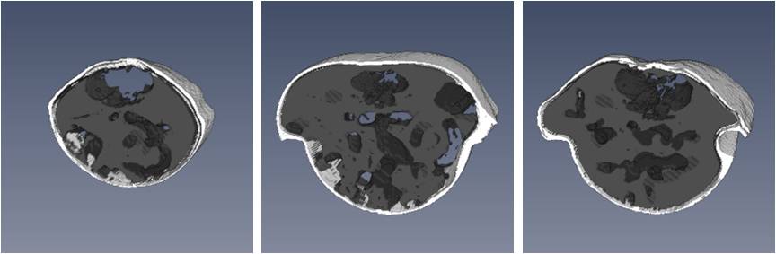
A novel segmental shape model is presented which separates visceral adipose tissue (VAT) from subcutaneous adipose tissue (SAT). With shape and distance constraints, it deforms a contour inwards from the skin to the muscle wall and separates the connecting adipose tissues in an ob/ob mouse. The fat tissues are segmented by the adaptive fuzzy C means method to compensate for intensity variation in adipose images. The results are obtained by logical operations applied on the extracted fat images and the separated adipose masks. Body Mask
In order to minimize the effect of inhomogeneity, we obtained the body mask through morphological operations on the edge images. Visceral MaskThe visceral mask is defined as the area inside the body mask that includes the visceral fat and excludes the subcutaneous fat. The contour of the visceral mask is called the visceral contour. In this step, we obtained the visceral mask by shrinking the body contour to the visceral contour.
Segmental Weight TemplateIn our application, because the ob/ob mice are too fat to easily fit into the holder, the extra fat tissues in bilateral regions usually fold the skin which decreases the shape similarities between the body and visceral contours. Therefore we designed a segmental weight template to include the capacity of adjusting the shape similarities along the contours.
Fat ExtractionThe adaptive fuzzy C means method (AFCM) was used here to extract fat tissues from muscle and other organs. During the standard fuzzy C means segmentation, the AFCM adaptively modifies the objective function to compensate for intensity inhomogeneity. The inhomogeneity field is decomposed from the raw image as a slow changing smooth surface and the corrected image has more equal intensities in fat tissues. Publication-Yang Tang, Priyank Sharma, Marvin D.Nelson, Simerly Richard, Rex A. Moats, Automatic Abdominal Fat Assessment in Obese Mice Using a Segmental Shape Model. Journal of Magnetic Resonance Imaging. 34 (4), pg. 866-873, 2011 |